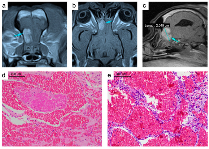
Figure 1.
(a–c) Magnetic resonance images of the tumor. The T1-weighted sequence used for the diagnosis confirming an intra-axial trabeculated mass (arrows) in the left olfactory bulb; transversal (a), coronal (b) and sagittal (c) images. (d,e) Hematoxylin and eosin-stained tumor biopsy. Multiple thin vessels replete with blood and thromboses (d). Scale bar: 100 μm.

