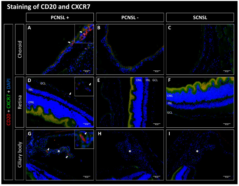
Figure 3.
Fluorescence microscopy of CD20 (red) and CXCR7 (green) double staining of the eyes in the PDX PCNSL vs. PDX SCNSL group. (A–C) show recordings of the choroid, (D–F) of the retina and (G–I) of the ciliary body (marked with *). (A,D,G) show the choroids, retinas, and ciliary bodies infiltrated with CD20-positive primary CNS lymphoma cells (PCNSL+) (arrows). Hardly any CXCR7-positive staining was found. No CD20+ cells were found in the PCNSL negative (PCNSL-) group (B,E,H) and in the SCNSL group (C,E,I). In the PCNSL- and SCNSL groups, CXCR7 stained faintly in the choroids (B,C), weakly to moderately in the retinas (E,F) and moderately in the ciliary body (H,I). ONL = outer nuclear layer, INL = inner nuclear layer, GCL = ganglion cell layer; scale bar 50 µm.

