Figure 5.
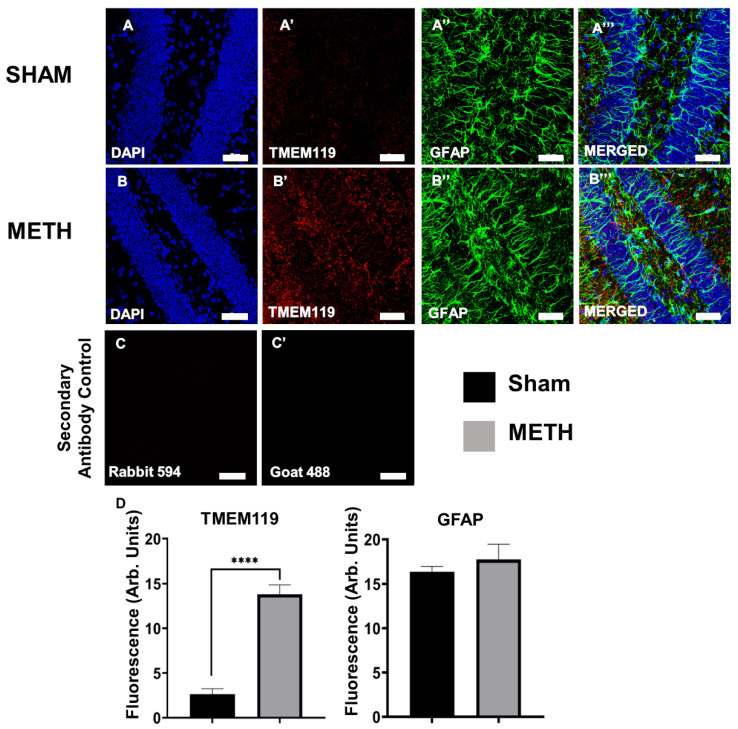
Acute METH exposure induces gliosis within the brain. Brain hippocampus cross-sections of sham-treated (A,A’,A’’,A’’’) and METH-treated (B,B’,B’’,B’’’) mice. (A,B) DAPI nuclei staining, (A’,B’) astrocytes labeled with anti-GFAP antibody, (A’’,B’’) microglial cells labeled with anti-TMEM119 antibody, and (A’’’,B’’’) merged images of DAPI, GFAP and TMEM119. Coronal sections; 20 μm; bregma −1.82 mm. Images captured at 40×, scale bar: 50 μm. (C,C’) Secondary Antibody Control. (D) Statistical analysis of Fluorescence (Arb. Units). No significant differences were noted between the sham- and METH-treated mice in GFAP. Significant differences were shown between sham- and METH-treated mice in TMEM 119. Results are expressed as mean ± standard error of the mean (SEM), **** p < 0.001.

