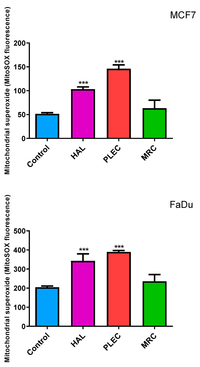
Figure 3.
Generation of mitochondrial superoxide in MCF7 and FaDu cells as detected with MitoSOX. The cells were exposed to HAL, PLEC and MRC and, after 24 h of incubation, MitoSOX Red fluorescence was measured using a microplate reader. Data are means ± SD (n = 3). *** (p < 0.001) as compared with control (untreated) cells.

