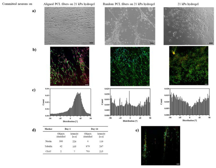
Figure 5.
Alignment of committed neurons on PCL-gelatin scaffolds. (a) Optical micrographs and (b) confocal micrographs−immunostained with tubulin, nestin, ChAT and DAPI (green, yellow, red, blue) −of neurons on aligned, randomly distributed PCL-gelatin scaffolds and 21 kPa gelatin hydrogels at day 14; (c) Histograms of alignment of neurites on fiber-hydrogel scaffolds, number of cells N = 100; (d) Intensity analysis of nestin and tubulin of neurons on aligned PCL-gelatin scaffolds, number of images N = 9; (e) Neuro-bundle arrangement on aligned PCL-gelatin scaffolds, immunostained with tubulin, nestin, ChAT and DAPI (green, yellow, red, blue) at day 14, all scale bars = 100 µm.

