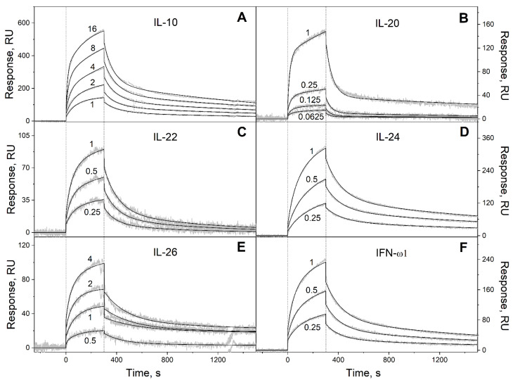
Figure 3.
Kinetics of the interaction of Ca2+-loaded S100P with specific four-helical cytokines of Interferons/IL-10 family shown in Table 2 at 25 °C, monitored by SPR spectroscopy using S100P as an analyte and the cytokines as a ligand immobilized on the sensor chip surface by amine coupling. (A): IL-10; (B): IL-20; (C): IL-22; (D): IL-24; (E): IL-26; (F): IFN-ω1. Buffer conditions: 10 mM HEPES, 150 mM NaCl, 1 mM CaCl2, 0.05% TWEEN 20, pH 7.4. The vertical dotted lines mark the association phase, followed by the dissociation phase. Molar analyte concentrations (µM) are indicated for the sensograms. The gray curves are experimental, while the black curves are theoretical, calculated according to the heterogeneous ligand model (1) (see Table 3).

