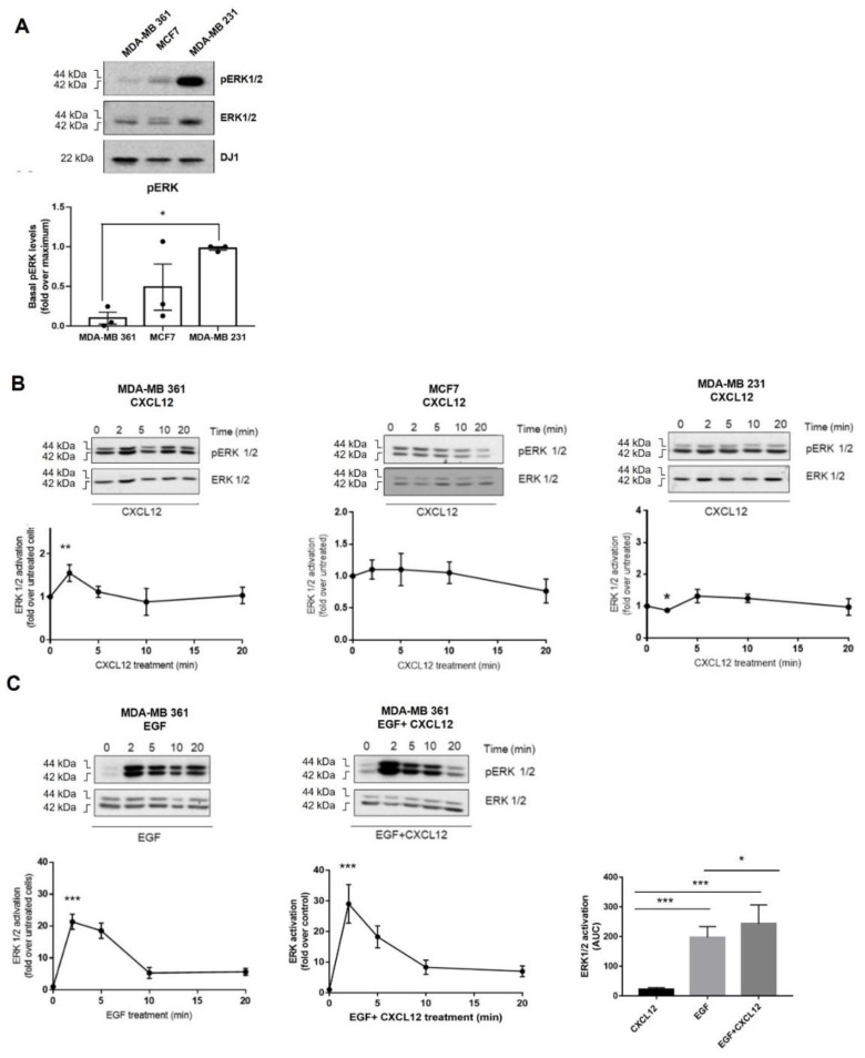
Figure 3.
(A) Basal ERK1/2 activation levels in distinct BC cell lines. (B) ERK pathway is poorly stimulated in MCF7 (ER+), MDA-MB361 (ER+ and HER2+) and MDA-MB231 (triple-negative) cells in response to exogenous CXCL12. Serum-starved cells were stimulated with 10 nM CXCL12 for the indicated times and ERK1/2 activation assessed as detailed in Methods. Activated phospho-ERK1/2 was normalized by total ERK levels and referenced as fold change over untreated cells. Representative blots are shown. * p < 0.05, or *** p < 0.001, comparing to non-stimulated cells. (C) Simultaneous activation of the CXCL12/CXCR4/ACKR3 and EGFR pathways fosters the ERK cascade stimulation pattern in MDA-MB-361 cells (ER+ and HER2+). Serum-starved cells were stimulated with 10 nM CXCL12, 100 ng/mL EGF or the combination of both ligands for the indicated times. ERK1/2 activation was assessed and plotted as above along with the area under the curve (AUC). Representative blots are shown. Data are mean ± SEM of 9–13 independent experiments. ** p < 0.01, or *** p < 0.001, comparing to non-stimulated cells.

