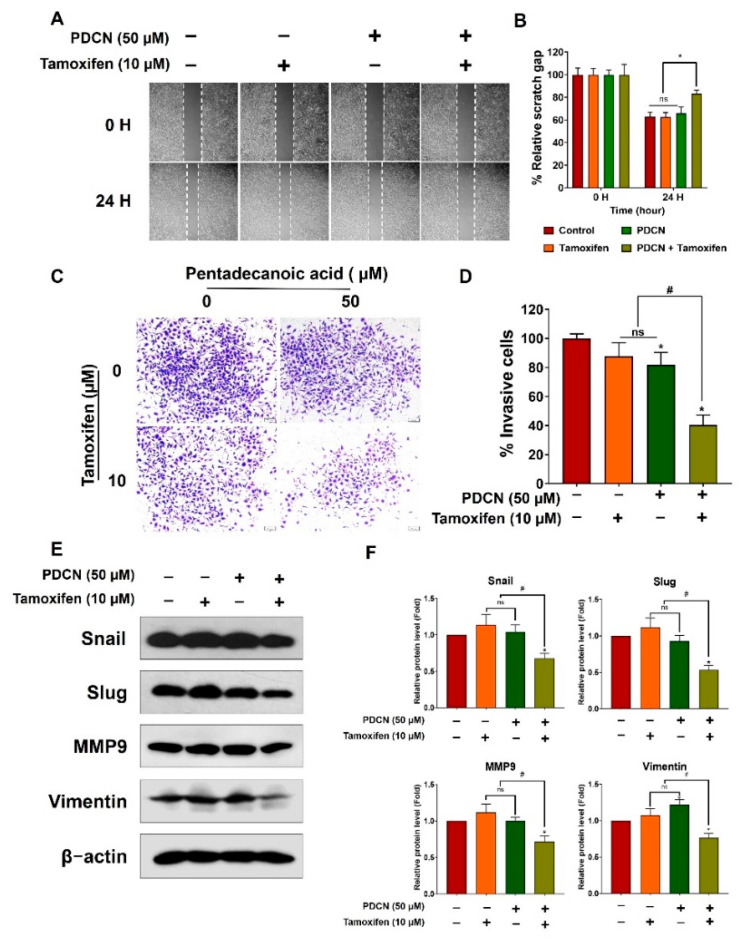
Figure 6.
Combined treatment with pentadecanoic acid and tamoxifen suppressed EMT. (A,B) Cell migration determined using the wound healing assay following treatment for 48 h in MCF−7/SC cells. (C,D) Invasive cells determined after pentadecanoic acid treatment alone or in combination with tamoxifen for 48 h. (E,F) Western blot analysis of epithelial–mesenchymal transition (EMT) markers in MCF−7/SC were performed after individual or combined treatment for 48 h. β−actin was used as a loading control. The asterisk (*) indicates p < 0.05 vs. the control. The hash mark (#) indicates p < 0.05 when comparing the combined vs. individual treatments. PDCN: pentadecanoic acid. Data are representative of three biologically independent experiments and values are shown in mean ± SD.

