Figure 4.
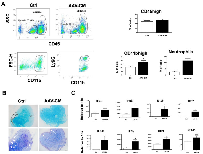
Intrathecal AAV-CM in mice with EAE induced the recruitment of myeloid cells, including neutrophils, to the CNS and reduced demyelination. (A) Flow cytometry profiles of brains of mice with EAE-treated with intrathecal AAV-CM or a control (ctrl). CD45high leukocyte populations are distinguished from CD45dim microglia. The proportion of CD45+ cells in the CNS remained similar in response to intrathecal AAV-CM treatment compared to the control EAE mice. Flow cytometric analysis revealed that the proportion of CD11bhigh and neutrophils were significantly increased upon intrathecal AAV-CM. n = 3–6 per group. (B) Images of lumbar spinal cord sections of mice with EAE stained with LFB and Cresyl violet. In the control group of mice, Cresyl violet staining showed cell infiltration into the parenchyma of the spinal cord (arrows), which was correlated with extensive loss of LFB (marked area) in corresponding areas. Mice with EAE that were treated with intrathecal AAV-CM showed cell accumulation in the meninges and reduced loss of LFB staining. Scale bar. 100 µm. (C) RT-qPCR analysis of brains showed IFNα, IFNβ, IFNγ, IL-10, IL-1β, IRF7 and IRF9 were significantly (STAT1, p < 0.0565) induced upon intrathecal AAV-CM treatment at 1 day post dose (n = 6–8). Data are presented as mean ± SEM. The results were analyzed using the two-tailed Mann–Whitney u-test. * p < 0.05, ** p < 0.01.

