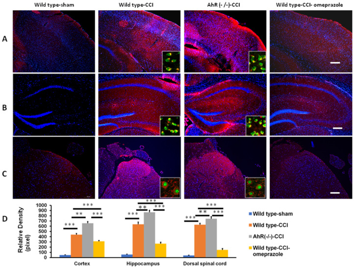
Figure 9.
The expression of inflammatory cytokines in the central nervous system in CCI subjected to the modulation of AhR. Either wild or AhR-deleted mouse received CCI injuries and then were subjected to omeprazole treatment. The somatosensory cortex, hippocampus, and dorsal spinal cord were obtained for analysis 28 days after injury (A) Expression of TNF-α in the somatosensory cortex in the different experimental groups. (B) Expression of TNF-α in the hippocampus in the different experimental groups. (C) Expression of TNF-α in the dorsal spinal cord in the different experimental groups. (D) Quantitative analysis of TNF-α in different groups. Magnification box indicates the imaging fusion of TNF-α and Neu-N (green). DAPI = blue; Bar length = 100 μm; N = 6; **: p < 0.01; ***: p < 0.01. Wild type-sham; Wild type-CCI; AhR (−/−)-CCI; Wild type-CCI-omeprazole: see text.

