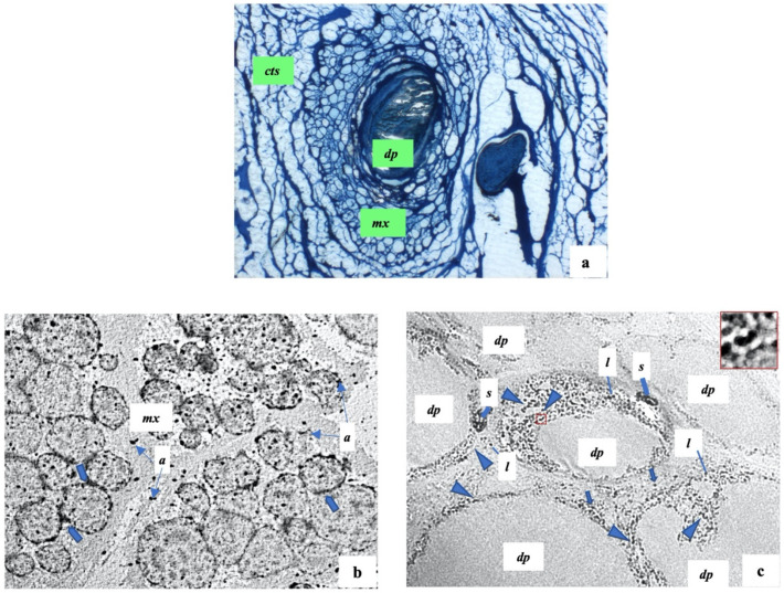
Figure 5.
Scalp hair follicles in alopecia areata. (a) A representative semithin section shows: a follicular papilla (dp) with abundant flocculent substance; a hair matrix (mx) with many cells not well organized; cts: connective tissue sheath. (b,c) The representative ultrastructural sections of hair matrix and follicular papilla respectively show: mx: hair matrix; a: apoptotic debris; thick arrows: beaded membrane of keratinocytes; dp: follicular papilla; head arrows: bacteria infiltrations; s: melanosome structures; l: lymphocytes; red square: area of inset magnification. (a) 200×; (b) 1750×; (c) 12,500×.

