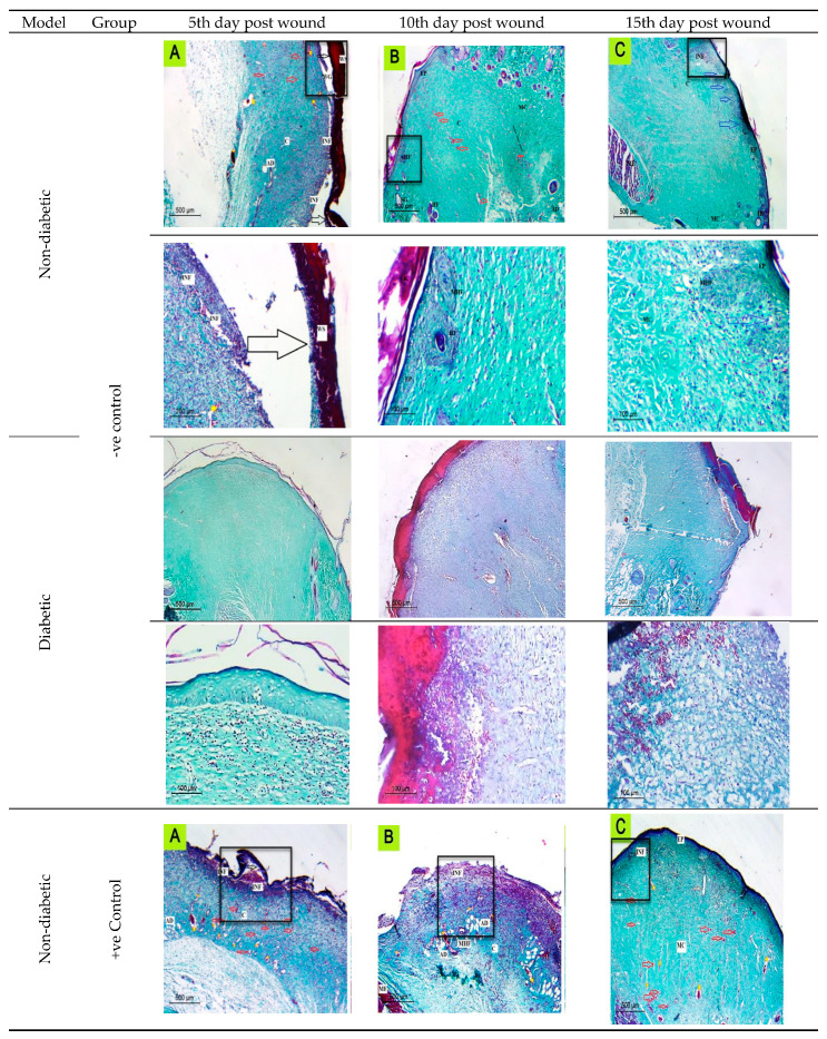
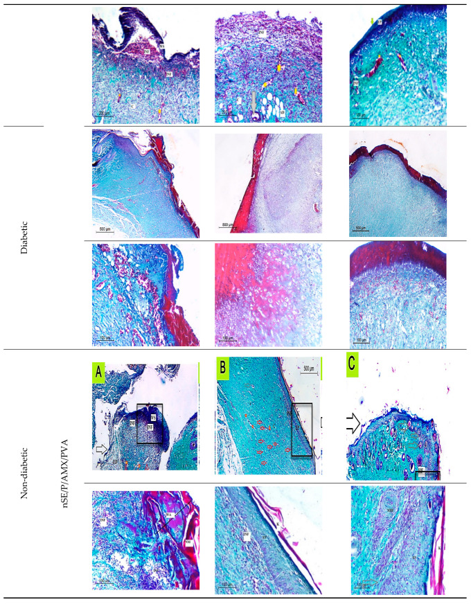
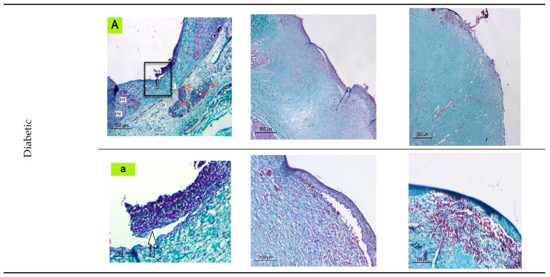
Figure 7.
The histological evaluation of skin wounds at different time intervals (5th, 10th, and 15th) indicated regression of the lesions with better epithelialization (blue arrows) and more effective re-organization of the dermis by collagen fiber maturation. Inflammatory cells (INF); Adipose connective tissue; blood vessels (red arrows); Immature collagen (IMC); mature collagen (MC); epidermis (EP); maturating hair follicle (MHF); Wound Gap (WG); wound area (WA); wound scab (WS) dilated blood vessels (Yellow Strikes); Muscle Fibers (MF); detached scabs (black arrows).


