Figure 1.
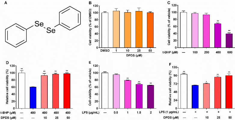
DPDS protected HBZY-1 cells against t-BHP-induced injury. The HBZY-1 cells were exposed to indicated concentrations of agents for 24 h, and cell viability was detected by a CCK-8 kit. (A) Chemical structure of DPDS. (B) Cell viability of HBZY-1 cells after treatment with indicated concentrations of DPDS for 24 h (n = 3). Reference: DMSO group. (C) t-BHP-induced cytotoxicity in HBZY-1 cells (n = 3). Reference: vehicle group. (D) Effects of DPDS on t-BHP-induced cytotoxicity (n = 6). Reference: the 400 μM t-BHP group. (E) LPS-induced cytotoxicity in HBZY-1 cells (n = 6). Reference: vehicle group. (F) Effects of DPDS on LPS-induced cytotoxicity (n = 6). Reference: the 1 μg/mL LPS group. Data are presented as means ± SEM. * p < 0.05, ** p < 0.01.

