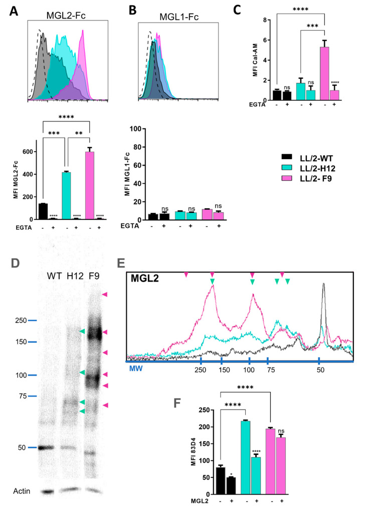
Figure 4.
MGL2 differently interacts with LL/2-Tn+ cell variants. (A,B) Recognition of LL/2 tumor cells with soluble MGL2-Fc (A) or MGL1-Fc (B) analyzed by flow cytometry. Bottom, Median fluorescence intensity (MFI), in the presence or absence of EGTA. (C) Binding of MGL2+ CHO cells to the LL/2 cell variants by solid phase assay. (D) Lectin blot analysis of whole cell lysates, membranes using MGL2-Fc. Green and violet arrows represent components recognized in LL/2-Tn+-H12 and LL/2-Tn+-F9 cells, respectively. (E) Densitometry of MGL2-Fc recognition of cell lysates by lectin blotting. (F) Anti-Tn 83D4 mAb staining of LL/2 cell lines, with or without pre-incubation with MGL2-Fc. Asterisks represent significant differences (* p < 0.05, ** p < 0.01, *** p < 0.001, **** p < 0.00001); ns: non-significant.

