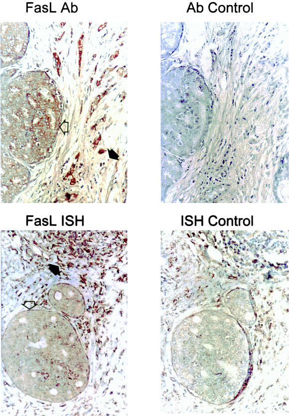
FIG. 1.
Human breast carcinomas express FasL. Immunoperoxidase staining with a FasL-specific rabbit polyclonal IgG antibody (FasL Ab) was performed with paraffin-embedded breast carcinoma sections. Slides were counterstained with hematoxylin. FasL-positive immunohistochemical staining (brown) is shown in a representative breast carcinoma (magnification, ×80). In addition to positive staining of the tumor island (open arrow), positive staining is also observed among isolated cells of lymphoid morphology (solid arrow), possibly representing FasL-expressing, activated T and NK cells. As a control for specificity of antibody detection, the FasL-immunizing peptide was included during primary antibody incubation (Ab control). Competitive displacement of staining by the soluble peptide immunogen confirms FasL specificity. Breast tumor expression of FasL mRNA was detected by in situ hybridization with a biotinylated FasL-specific riboprobe (FasL ISH). A positive brown hybridization signal is seen within a representative tumor island (open arrow) (magnification, ×80). FasL mRNA was also detected in cells within a lymphoid aggregate (solid arrow). In control sections for in situ hybridization (ISH control), the biotinylated sense control probe failed to hybridize, confirming the specificity of the FasL hybridization. These results are representative of 17 breast carcinomas.

