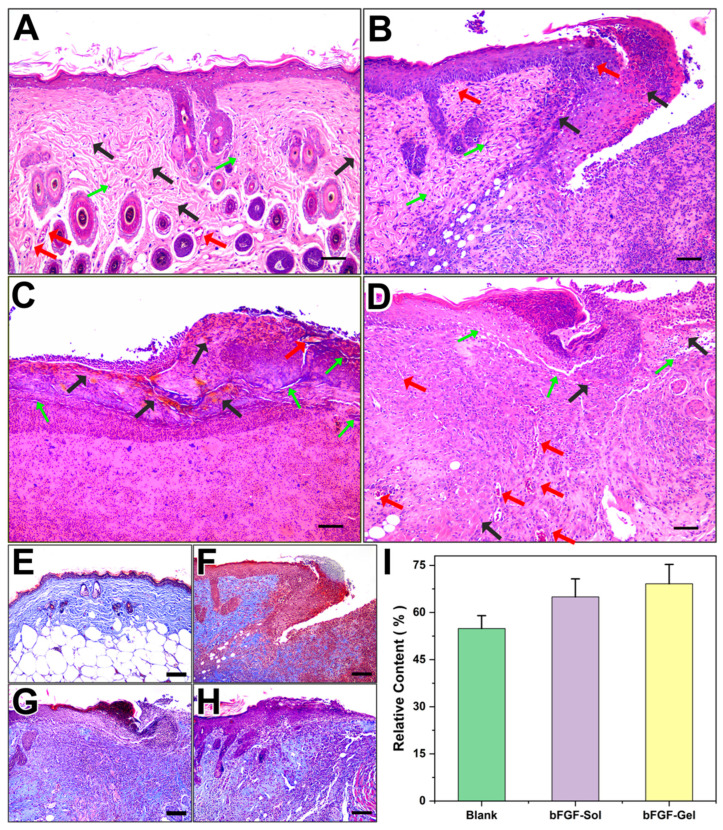
Figure 4.
Histological analysis of wounds on the dorsum of mice by H&E staining after treatment with no bFGF, spraying bFGF, and hydrogel dressing containing bFGF for up to 10 days. (A) normal skin, (B) Blank, (C) bFGF-Sol, (D) bFGF-Gel. Scale bar =100 μm. (Green arrows: fibroblasts; black arrows: collagenous fibers; Red arrows: blood capillaries.). Masson staining images from day 10 post-operation. (E) normal skin, (F) Blank, (G) bFGF, (H) bFGF-Gel, (I) relative content of collagen in different groups compared with normal skin. Scale bar =100 μm.

