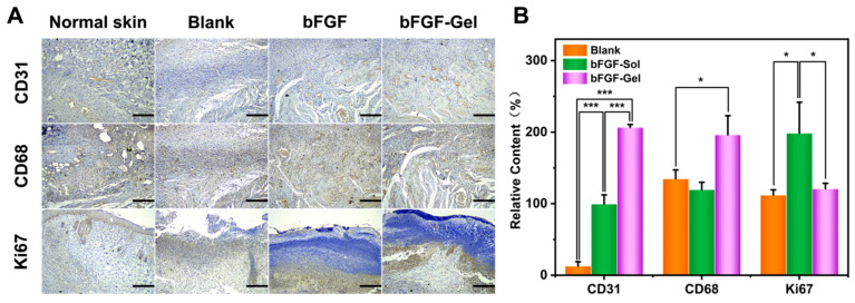
Figure 5.
Immunohistochemical analyses of wounds on the dorsum of mice by CD31, CD68, and Ki67 staining after 10 days. (A) Representative images of CD31, CD68, and Ki67 staining in groups of Normal skin, Blank, bFDF, and bFGF-Gel at day 10; Scale bar = 200 μm. (B) Semiquantitative results of CD31, CD68, and Ki67 staining. The data were represented as mean ± SD (n =3). * p < 0.05, *** p < 0.001. Scale bar = 200 μm.

