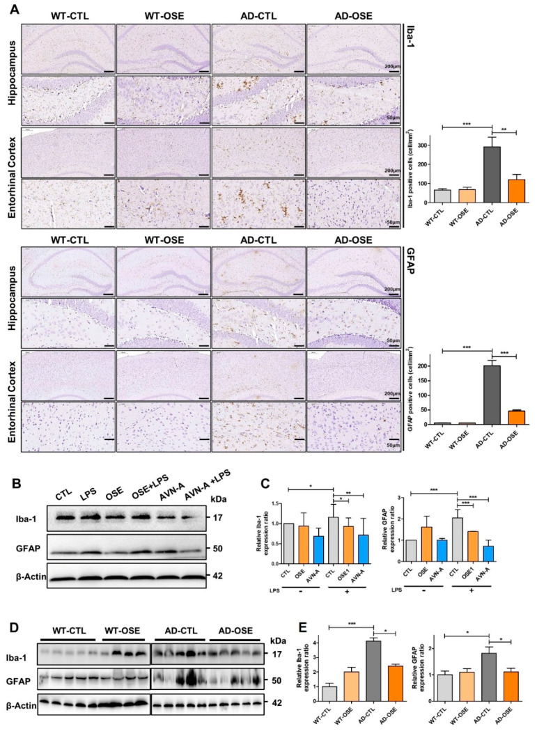
Figure 4.
Reduced reactive astrocytes and active microglia. (A) Immunohistochemistrical staining of GFAP and Iba1 within the brain sections’ hippocampus and entorhinal cortex in Tg-5xFAD AD and WT mice after exposure to OSE. (B,D) GFAP and Iba1 Western blotting in cell line and Tg-5xFAD AD mice after exposure to OSE and AVN-A. Actin is being utilized as the loading control. (C,E) Validation of differentially expressed proteins by Western-blot analysis. * p < 0.05, ** p < 0.01, *** p < 0.001.

