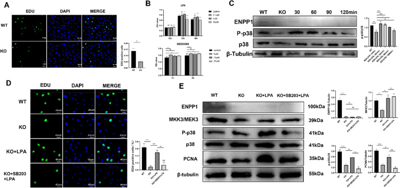
Fig. 4.
Lysophosphatidic acid (LPA) partially rescued the impacted proliferation of Enpp1 KO pre-osteoblasts via the MKK3/p38 MAPK/PCNA pathway A EdU immunostaining of Pobs from WT and Enpp1 KO suckling mice. EdU-positive cells were quantified (n = 3). B Cell viability measurement of Pobs incubated with LPA under a series of concentrations (0–10 μM) and times (12–48 h) or incubated with SB203580 (0–20 µM, 1 h, and 2 h) (n = 8). C Immunoblot showed the expression of phosphorylated p38 MAPK/p38 MAPK of WT and Enpp1 KO Pobs at different time gradients (0, 30, 60, 90, and 120 min) under LPA stimulation at 1 uM concentration (n = 3). D Pobs of WT and Enpp1 KO groups were incubated in the presence of LPA (1 μM) or SB203580 (10 μM; added 2 h prior to LPA addition) followed by LPA (1 µM) for 48 h. Cells were then stained for EdU immunostaining and visualized using confocal microscopy. Fluorescence intensity was quantified using Image-Pro Plus 6.0 (Media Cybernetics). Three different areas per chamber were measured. E Serum-starved Pobs were treated with LPA in the absence or presence of SB203580 (10 μM) that was added 2 h prior to LPA addition. MKK3/p38 MAPK/PCNA pathway expression was monitored using western blot. One representative blot for each protein and the densitometric analysis (mean + SD) from three independent experiments are presented. To improve clarity and conciseness, blots are cropped to the location of the target protein band. (Results are presented as the mean ± SD, *p < 0.05, **p < 0.01, ***p < 0.001; one-way analysis of variance with Bonferroni correction)

