Fig. 1.
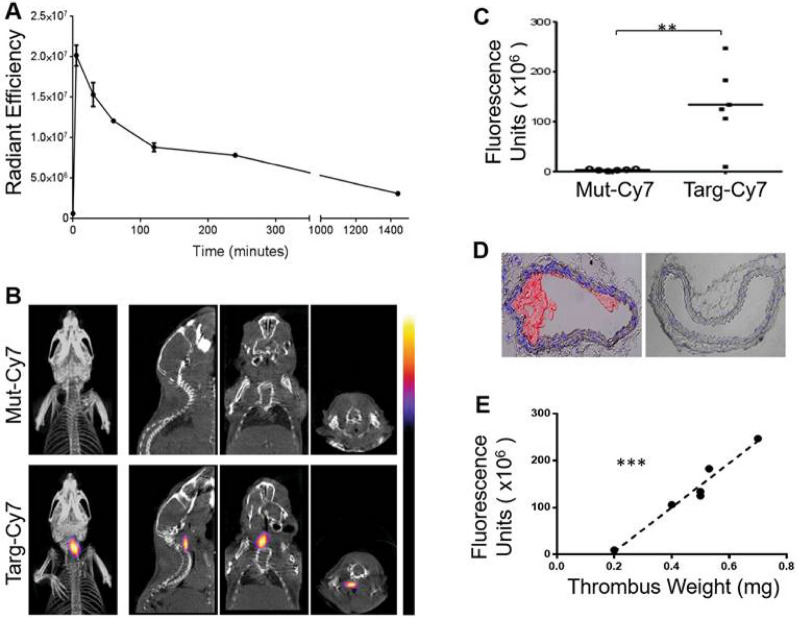
InSyTe FLECT/CT imaging of mice with left carotid ferric chloride–induced thrombosis using a targeted NIR fluorescent fluorophore. A NIR fluorescence signal of Targ-Cy7 in collected blood samples as determined by IVIS® Lumina to determine in vivo circulatory half-life before imaging using FLECT-CT. B FLECT-CT scans of mice with left carotid thrombosis showing selective binding of targeting fluoroprobe (Targ-Cy7; bottom panel), compared to mutated control (Mut-Cy7; top panel). C NIR fluorescence units of Targ-Cy7 and Mut-Cy7. D Representative images of ferric chloride–injured carotid artery (left) and contralateral non-injured carotid artery (right), where nuclear stain (DAPI) is blue and platelet-specific (CD41-allophycocyanin) is red. E Further analysis of detected signal in each mouse shows a strongly significant correlation to the weight of its ex vivo thrombus. Adapted with permission from [57].
Copyright 2017 Ivyspring

