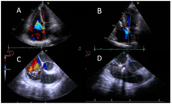
Figure 1.
Echocardiographic pictures of successful TR reduction after TEER. Transthoracic echocardiographic four-chamber view at baseline (A) and at discharge (B) after implantation of one TriClip XT. Transoesophageal echocardiographic right ventricle inflow/outflow view showing severe tricuspid regurgitation mainly originating from septal and anterior leaflet at baseline (C) and traces of regurgitation after implantation of one device (D).

