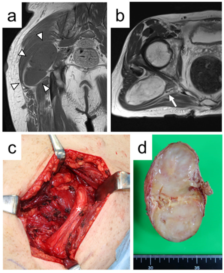
Figure 1.
A 55-year-old male with nodular plexiform neurofibroma of the right buttock (Case 7). (a) Coronal T1-weighted magnetic resonance image shows an intramuscular tumor with homogeneous iso-signal intensity compared with skeletal muscle (arrowheads). (b) Axial T2-weighted magnetic resonance image reveals that the tumor between the piriformis muscle and gluteus maximus muscle is adjacent to the sciatic nerve (arrow). (c) An intraoperative photograph shows that the tumor originated from the branch of the sciatic nerve (asterisk) to the gluteus maximus muscle and was treated with en bloc resection. (d) The en bloc resected specimen reveals a yellowish-white tumor on gross finding (Tumor 10).

