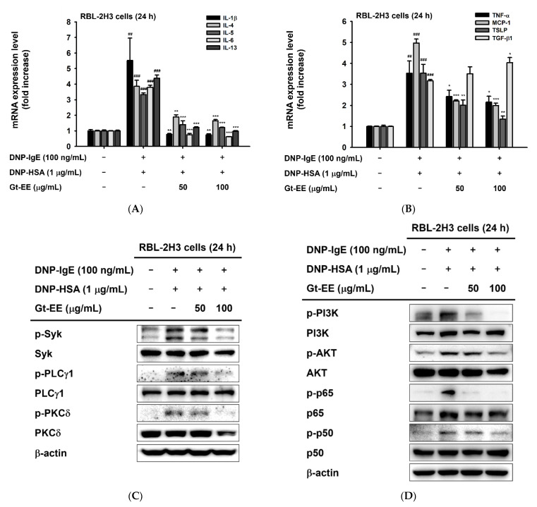
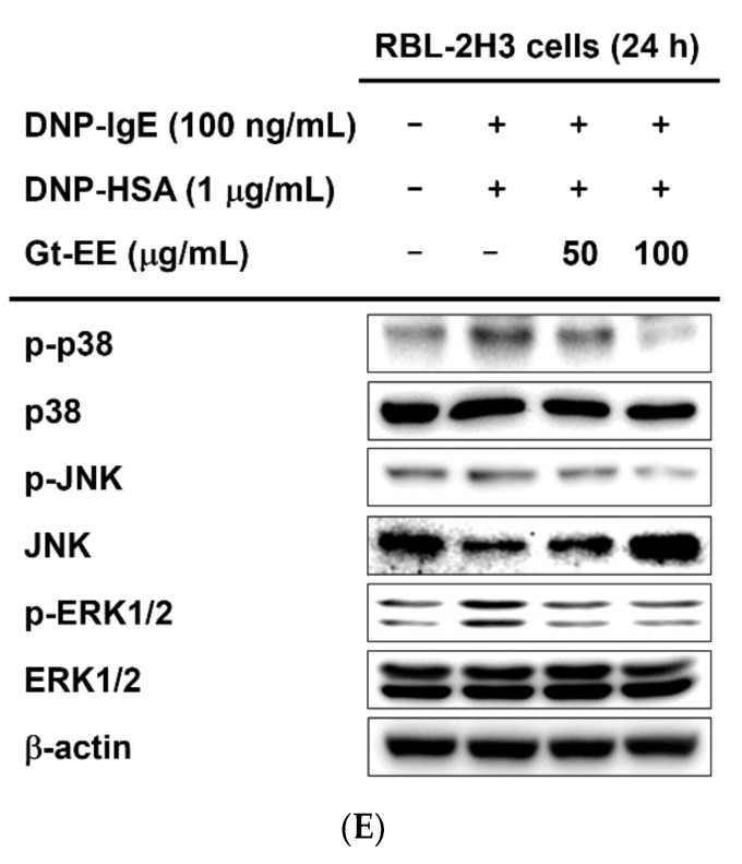
Figure 2.
Gt-EE suppressed the mRNA expression of allergic cytokines and activation of the IgE–FcεRI signaling pathway. (A,B) The mRNA expression levels of IL-1β, IL-4, IL-5, IL-6, IL-13, TNF-α, MCP-1, TSLP, and TGF-β1 in IgE-stimulated RBL-2H3 cells were investigated using real-time PCR. IgE-sensitized RBL-2H3 cells were treated with Gt-EE for 30 min and then challenged with DNP-HAS for 24 h. (C–E) The total or phosphorylated forms of Syk, PLCγ1, PKCδ, PI3K, AKT, NF-κB p65, NF-κB p50, p38, JNK, and ERK1/2 in IgE-stimulated RBL-2H3 cells were detected using an immunoblotting analysis. IgE-sensitized RBL-2H3 cells were treated with Gt-EE for 30 min and then stimulated with DNP-HAS for 24 h. (A,B) The results are expressed as mean ± standard deviation. ## p < 0.01, ### p < 0.001 compared with the normal group, and * p < 0.05, ** p < 0.01, *** p < 0.001 compared with the control group.


