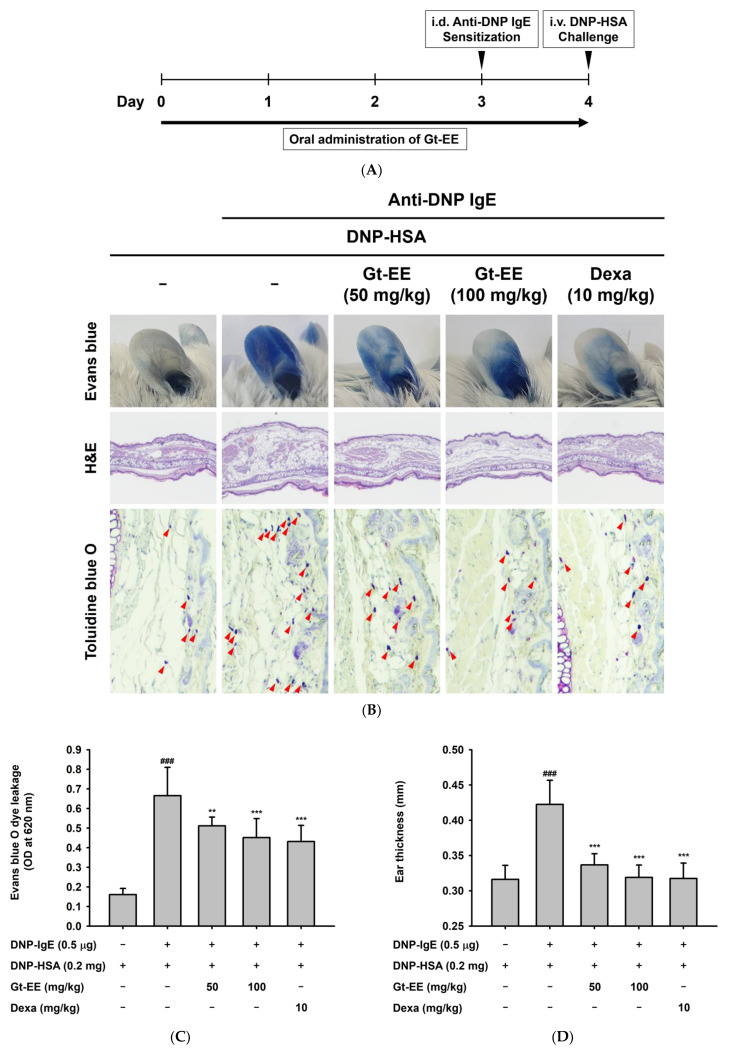
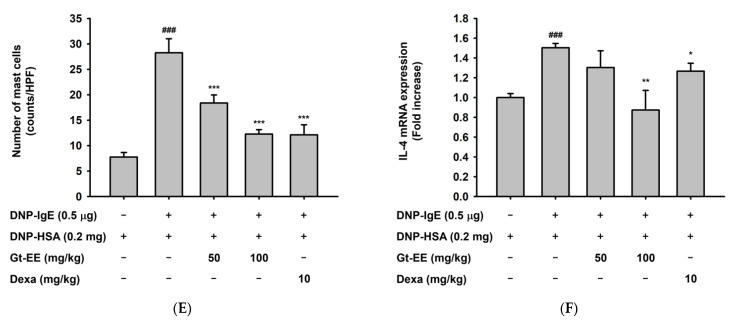
Figure 3.
Effects of Gt-EE on PCA. (A) Experimental procedure for induction of PCA. Anti-DNP IgE and DNP-HSA were administered to the ears of BALB/c mice to induce PCA, and then the ears were stained with Evans blue dye. Gt-EE was orally administered every day for 5 days before PCA induction. Dexamethasone was used as the positive control. After euthanasia, the ears were dissected and soaked in formamide for extravasation of the Evans blue dye or fixed for histopathological analysis; (B) photographs of the Evans blue–stained ears and ear tissues stained with hematoxylin and eosin (H&E) or toluidine blue O. Histopathological variations and alterations in the mast cell counts due to IgE–antigen induction were assessed using H&E and toluidine blue O staining, respectively. The red arrows indicate toluidine blue O–stained mast cells; (C) absorbance of Evans blue extravasated from the mouse ears. Absorbance was measured at 620 nm. (D) The change in ear thickness was investigated using dial thickness gauges (PEACOCK, Japan). (E) The number of mast cells in the ear tissue was counted under a microscope at 400× magnification. (F) The mRNA expression of IL-4 in the mouse ears was measured using real-time PCR. After euthanasia, the ears were dissected and ground, and then total RNA was isolated for real-time PCR. (C–F) The results are presented as the mean ± standard deviation. ### p < 0.001 compared with the normal group, and * p < 0.05, ** p < 0.01, *** p < 0.001 compared with the control group.


