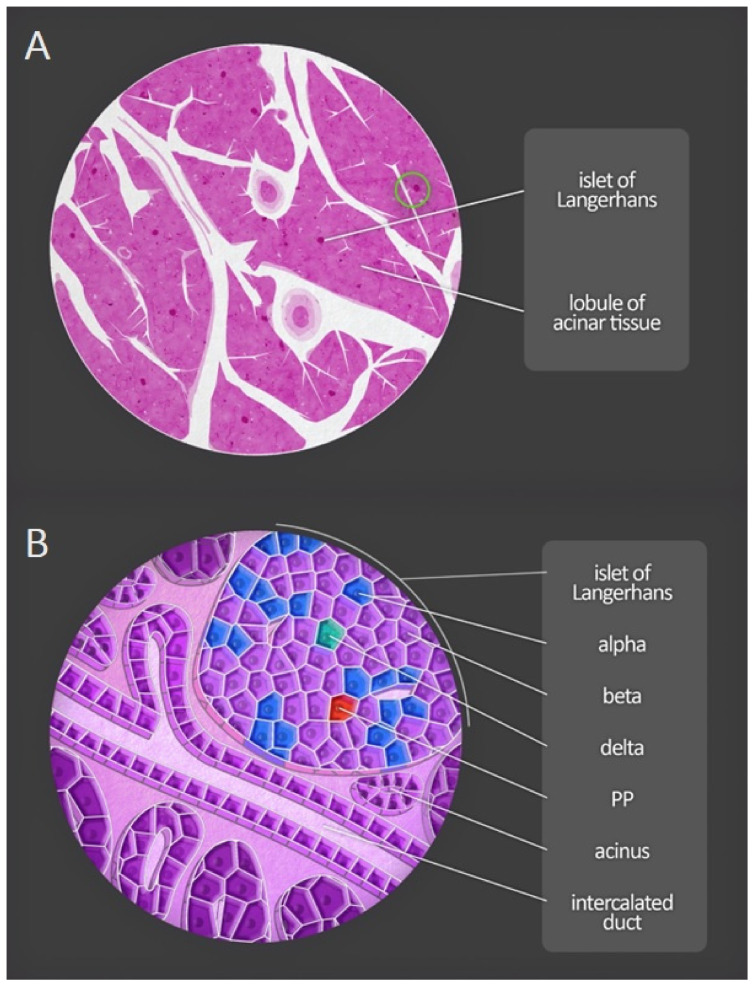
Figure 4.
Microscopic anatomy of the human pancreas. (A) Magnification of a portion of the pancreas shows both lobules and pancreatic islets. (B) At greater magnification, acini and excretory ducts are visible. Different types of endocrine cells composing the pancreatic islets can be distinguished by immunofluorescent staining. PP—pancreatic polypeptide cells.

