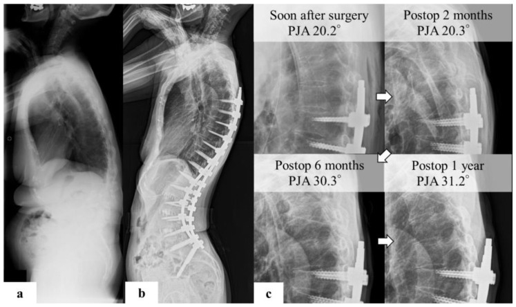
Figure 4.
(a) Representative case of PJK preoperative standing X-ray images indicated the following spinal parameters: C7SVA, 86 mm; TK, 20°; LL, 20°; PT, 30°; PI, 53°; TPA, 32°; and PI–LL, 23°. (b) postoperative images show that the PJA was 20.2. (c) PJA developed 6 months postoperatively. The PJA was 31.2° in the first year postoperatively, and the change was 11.2°. This patient complained of implant prominence and pain in the proximal junctional area, but refused reoperation.

