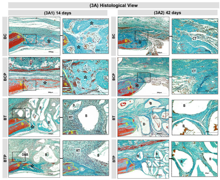
Figure 3.
Histological views (3A) in calvaria defects created in the animals. BC and BCP treated with blood clot and BT and BTP treated with demineralized bovine bone plus fibrin sealant at experimental periods 14 and 42 days (3A1,3A2). At 14 days: BC and BCP groups showed bone growth (asterisks) adjacent to the defect border (b) and on the dura-mater surface. The BT and BTP groups showed a discrete bone growth (asterisks) on the border and the defect was filled by graft particles (demineralized bovine bone) surrounded by a reactional tissue (RT) containing some inflammatory cells (3A1). At 42 days: BC and BCP both groups showed similar bone formation with gradual increase in thickness of trabeculae, leading to a compact structure limited to the defect border. Closure of large part of the defect by fibrous connective tissue (CT). In BT and BTP groups, the graft particles and inflammatory process decreased but did not disappear and only small bone formation was present on the lesion border (3A2). (Masson’s trichrome; original magnification 10×; bar = 500 µm and Insets, magnified images 40×; bar = 100 µm).

