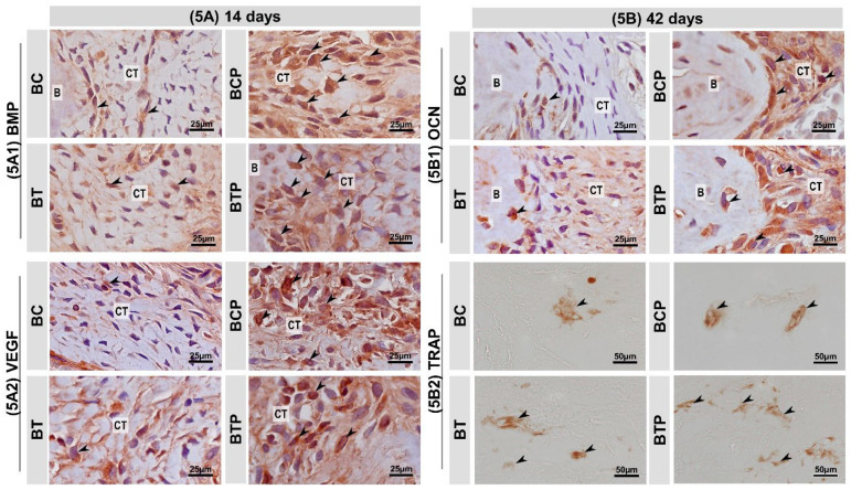
Figure 5.
(5A,5B) Histological sections showing the appearance representative of immunolabeling after 14 and 42 days respectively in experimental groups BC and BCP treated with blood clot biostimulated by laser or not and BT and BTP treated with demineralized bovine bone plus fibrin sealant biostimulated by laser or not. The black arrows point to places where the brown spot marks the proteins: (5A1) immunolabeling for bone morphogenetic protein (BMP—2) in bone defects at 14 days. BMP—2/4—positive cells (arrows); connective tissue (CT). (5A2) Immunomarking pattern for vascular endothelial growth factor (VEGF) in bone defect at 14 days. VEGF—positive cells (arrows); connective tissue (CT). (5B1) Immunomarking pattern for osteocalcin (OCN) in bone defect at 42 days. OCN—positive cells (arrows); connective tissue (CT) and bone tissue (B). (5B2) Immunomarking pattern for tartrate-resistant acid phosphatase (TRAP) in bone defect at 42 days. TRAP—positive cells (arrows). Harris’ hematoxylin counterstaining (Scale bars: 25 μm and 50 μm; original magnification: 40× and 100×, respectively).

