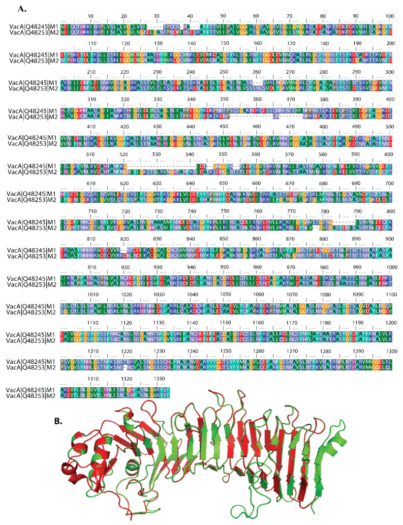
Figure 1.
Sequence and structure of VacA isoform. (A) Comparison of amino acid sequences of M1 and M2 isoforms of VacA protein of Helicobacter pylori. Corresponding accession nos. Q48245 (H. pylori strain 60190, m1 VacA) and Q48253 (strain Tx30a, m2 VacA). Amino acid color code generated by ClustalW within BioEdit software was used. Consensus residues ("Clustal cons" line) were generated by ClustalW. The numbers at the beginning and end of each line are for reference only and do not correspond to the original numbers of individual amino acid sequences recorded in published reports. Hyphens indicate gaps in the alignment at that position. (B) The ribbon image shows the superimposition of the I-TASSER modelled m1 protein (green color) with m2 protein (red color).

