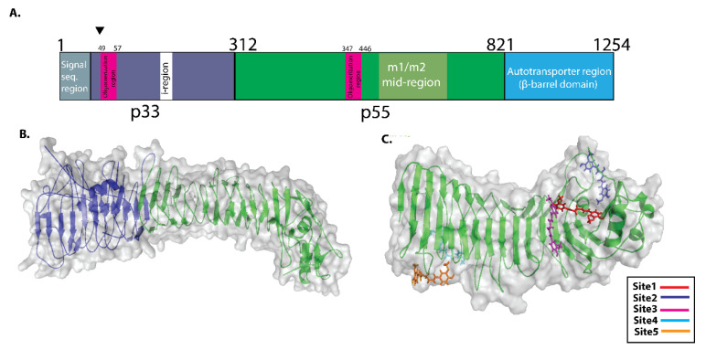
Figure 2.
Schematic depiction of VacA protein structure and potential binding sites for Curcumin. (A) p33 domain of VacA protein (1–311 amino acid sequence region) (shown in blue colour). The p55 domain of VacA (312–821 amino acid sequence region) (shown in green colour). VacA p33 region includes an initial signal sequence, an oligomerization region in the middle, and an intermediate (“i”) region at the end. The mid-region amino acid sequences of the p55 domain of VacA protein define m1 and m2 isoforms (B) The VacA p33 and p55 are three-dimensional structures. (The p33 and p55 domains are shown in blue and green colors, respectively. (C) COACH server predicted potential binding site present in p55 domain of VacA protein. Site1, site 2, site 3, site 4, and site 5 are shown in different colors.

