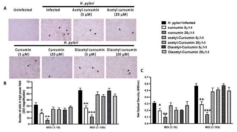
Figure 6.
Effect of curcumin and non-oxidizable derivatives on H. pylori mediated VacA vacuolation activity in AGS cells (* p value < 0.05; ** p value < 0.01; *** p value < 0.001 vs H. pylori infected). Data are expressed as means SEM, n = 3 independent replicates. (A) Light microscopy images showed uninfected AGS cells. The black arrow indicated the vacuolation region in H.pylori-infected AGS cells pretreated with curcumin and non-oxidizable curcumin. (B) The histogram graph represented the inhibition of vacuolation activity in H. pylori-infected AGS cells at two MOI (10:1, 100:1) and pretreated with curcumin and non-oxidizable curcumin. (C) The histogram graph showed neutral red uptake by AGS cells at OD 540 nm in H. pylori-infected AGS cells at two MOI (10:1, 100:1) and pretreated with curcumin and non-oxidizable curcumin. (* p value < 0.05; ** p value < 0.01; *** p value < 0.001 vs. H. pylori infected). Data are expressed as means SEM, n = 3 independent replicates.

