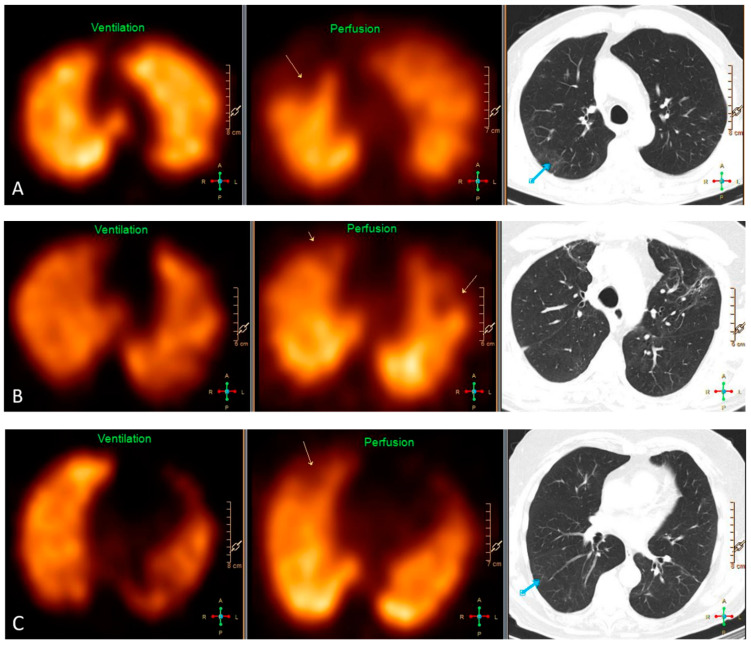
Figure 2.
Representative findings on VQ SPECT and HRCT of three patients. (A) Pulmonary embolism in the right upper lobe causing a segmental mismatched perfusion defect on SPECT (yellow arrow) without any abnormality in the same area on HRCT. The blue arrow depicts ground-glass opacities dorsally in the right upper lobe without any defect on SPECT. (B) HRCT shows signs of fibrosis in the upper lobes causing partially mismatched subsegmental perfusion defects on SPECT (yellow arrows). (C) Pulmonary embolism in the right upper lobe causing a subsegmental mismatched perfusion defect on SPECT (yellow arrow) without any abnormality on HRCT in the same area. The blue arrow depicts discrete ground-glass opacities and signs of hypoventilation dorsally in the upper part of the right lower lobe.

