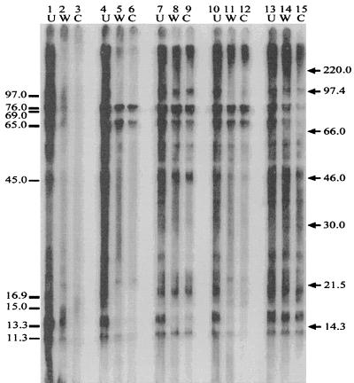
FIG. 4.
Immunoabsorption. Cat serum (cat 40) that was previously absorbed with whole cells (W) and cell lysates (C) of B. henselae LSU16 (lanes 2 and 3), B. henselae Houston-1 (lanes 5 and 6), B. clarridgeiae (lanes 8 and 9), B. quintana (lanes 11 and 12), or B. bronchiseptica, P. multocida, and E. coli (lanes 14 and 15) is shown. Unabsorbed serum (U) (lanes 1, 4, 7, 10, and 13) from cat 40 (B. henselae LSU16 infected) was treated in the same manner as the absorbed sera. The sera were allowed to react with B. henselae LSU16 antigen. Bartonella-specific antigens which were focused upon in this experiment are indicated on the left (in kilodaltons). Molecular mass markers are shown on the right (in kilodaltons).

