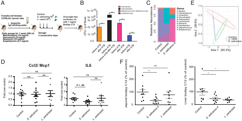
Fig. 5.
Duodenal isolates of oropharyngeal bacteria lead to decreased lipid absorption in vivo. (A) Schema of the experimental setup of the mouse experiments performed. (B) CFU for different groups of bacteria in the feces of mice prior to and after treatment with the antibiotic mixture for 7 consecutive days. (C) Average relative abundance of bacterial genera in the three treatment groups. (D) Expression of proinflammatory genes in the small intestine of mice overexposed to either S. salivarius or L. paracasei (control bacterium). Values are normalized to the geometric mean of the three housekeeping genes tbp, b2m, and gapdh. (E) PCoA (Principal Coordinate Analysis) on the Bray–Curtis index of the 16S amplicon data of the three treatment groups on ASV level; 95% CIs of the beta-dispersion of a given sample group are indicated with two ellipses. (F) BODIPY C12 absorption in the jejunum and liver of mice overexposed to S. salivarius or L. paracasei. Groups are compared using the Mann–Whitney U test. ATB, antibiotics. ns, P > 0.05. *P < 0.05; **P < 0.01; ***P < 0.001.

