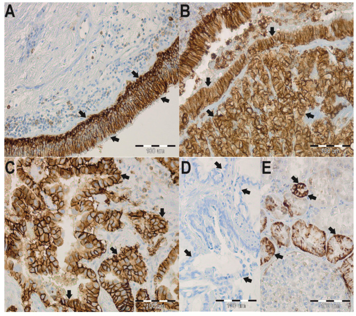
Figure 1.
Immunohistochemistry (IHC) staining of aquaporin 3 (AQP3) in paraffin-embedded sections of the lung adenocarcinoma (ADC) from humans. Anti-AQP3 antibody labels in non-neoplastic lung tissues. Arrows indicate the localization of the basolateral plasma membranes of surface epithelium in whole plasma membranes of basal cells of the bronchus (A). Anti-AQP3 antibody labels in the cancer lung tissues. Arrows indicate the localization of AQP3 in the cytoplasmic membrane in the cancer cells (B,C). No staining was observed when isotype-specific immunoglobulins from non-immunized rabbits were used instead of the primary antibody (negative control) (D). Immunoperoxidase labeling of AQP3 from the human kidney (positive control) (E). The labeling is seen in the basolateral plasma membrane of the renal collecting ducts. The blue color represents a positive staining for the hematoxylin counterstain, and the brown color represents positive AQP3 stain.

