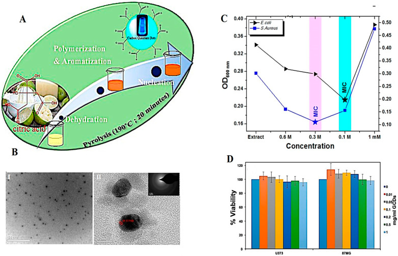
Figure 2.
(A) The preparative process of CDs using C. Limetta waste pulp. (B) (I) High-resolution transmission electron microscopy (HR-TEM) image (scale bar: 100 nm), (II) lattice fringe analysis (scale bar: 10 nm). (C) OD600 measurements of bacterial cultures incubated with CDs with different mM for analyzing minimum inhibitory concentration (MIC). (D) The viability analysis of cells treated with CDs using CKK8 assay. Reproduced with permission from Ref [37]. Copyright 2018 American Chemical Society.

