Figure 3.

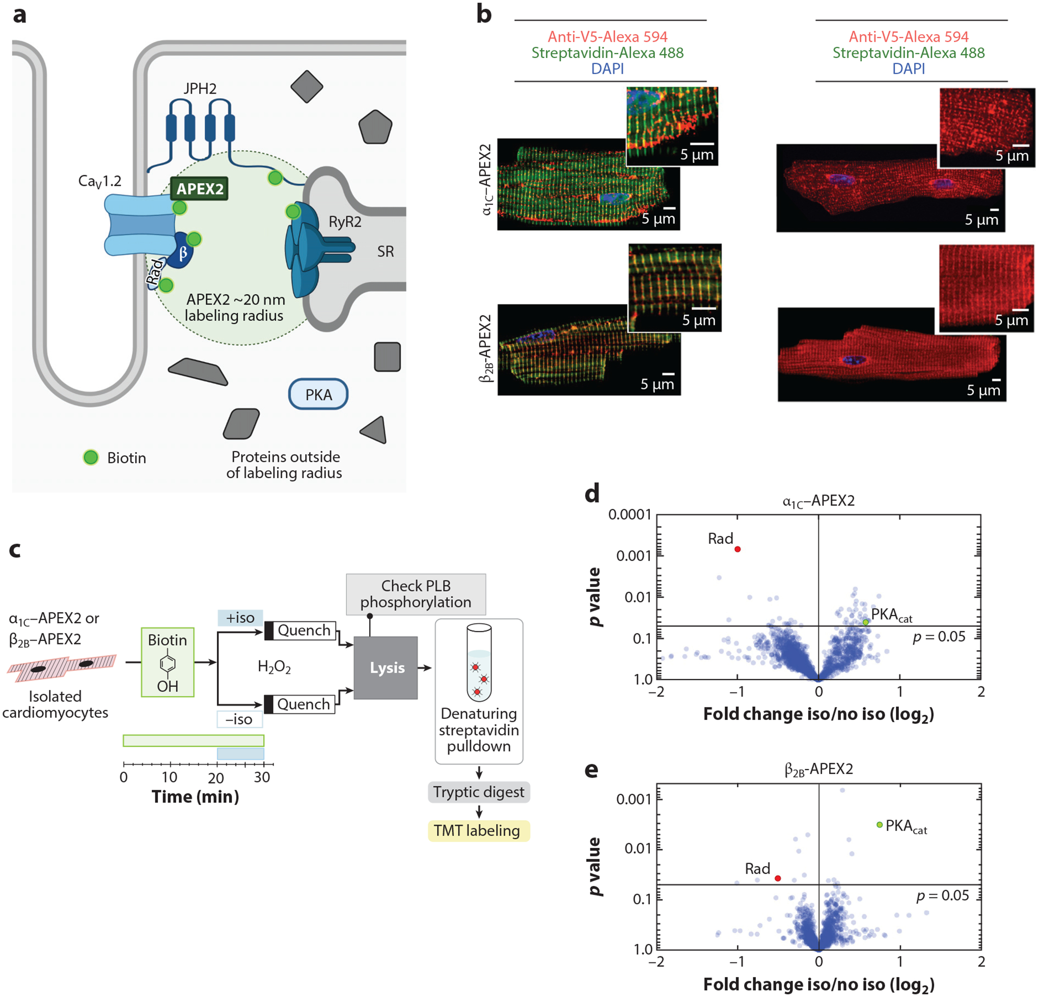
Proximity labeling using APEX2 to identify the mechanism underlying adrenergic agonist-induced augmentation of Ca2+ current. (a) Schematic depicting localization of APEX2-conjugated CaV1.2 channels in the dyadic space of cardiomyocytes. Panel a adapted from image created with BioRender.com. (b) Immunofluorescence of cardiomyocytes isolated from α1C-APEX2- and β2B-APEX2-expressing mice exposed to biotin-phenol and H2O2 or no H2O2. Nuclear labeling with DAPI stain. (c) Schematic of workflow for isolated cardiomyocytes. (d–e) Volcano plots of fold-change for relative protein quantification by tandem mass tag mass spectrometry of α1C-APEX2 and β2B-APEX2 samples. Non-adjusted unpaired two-tailed t-test. Rad (red dots) is reduced and PKA catalytic subunit (PKAcat; green dots) is increased. Panels b–e adapted from Reference 68. Abbreviations: APEX2, ascorbate peroxidase 2; JPH2, junctophilin 2; PLB, phospholamban; Rad, Ras associated with diabetes; RyR2, ryanodine receptor 2; SR, sarcoplasmic reticulum; TMT, tandem mass tag.
