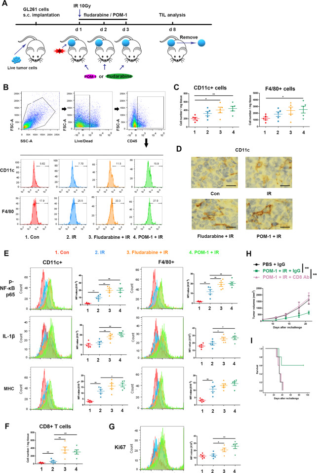
Fig. 4. STAT1 or CD39 inhibition enhanced inflammasome activation of mononuclear phagocyte system in TME following IR.
Subcutaneous tumors of C57BL/6 J mice were irradiated after mice were administrated with PBS, fludarabine, or POM-1, then were removed to prepare single-cell suspensions. Cells were stained for surface markers, then fixed and permeabilized for intracellular staining. Flow cytometry was used for markers measurement. A The schedule of treatment for mice is showed. B Representative plots showing the general gating strategy to delimit the positive CD45 cells from tumor tissue and positive CD11c and F4/80 cells in CD45+ T cells. C Positive CD11c and F4/80 cell number. D Positive CD11c cells were analyzed using IHC, and Images were captured with a light microscope (×400). Scale bars correspond to 20 μm. E The levels of phospho-NF-κB p65, IL-1β, and MHC in CD11c + or F4/80+ cells. F Positive CD8 cell number. G The levels of Ki67 in CD8 + cells. H, I The mice were removed from the untreated or IR + POM-1-treated tumors. were injected with live GL261s cells on the other side, and administrated with or without CD8 antibody. H the rechallenged tumor size was measured. I The survival of mice was recorded. *P < 0.05, **P < 0.01.

