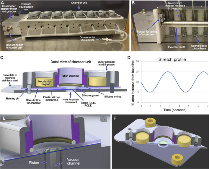
FIGURE 1.
An image of a stretch device with eight chamber units for mounting of lung slices (A). The unit is placed in a conventional tissue culture incubator and controlled by a programmable control unit placed outside the incubator. A piston is located underneath each chamber unit and produces the stretch movement by pushing the elastic membrane and the lung slice upwards. A vacuum line is connected to ensure contact between the elastic membranes and the driving pistons during the return phase. An image of part of the underside of a stretch device shows the off-center mounted shaft that drives the movement of the piston (B). A sensor feeds information on the achieved movement to the control unit as part of a closed loop-control system. A schematic outline of the design of the chamber units (C). Each chamber unit is held together by the attraction between the neodymium magnets and the metallic baseplate. Additional magnets mounted in the baseplate (B) help keep the chambers together when mounted on the stretch device. The movement of the shaft and thereby the achieved stretch can be programmed to emulate different breathing patterns, a simple sinusoidal profile has been used in the current study (D). Close-ups of the well design (E,F).

