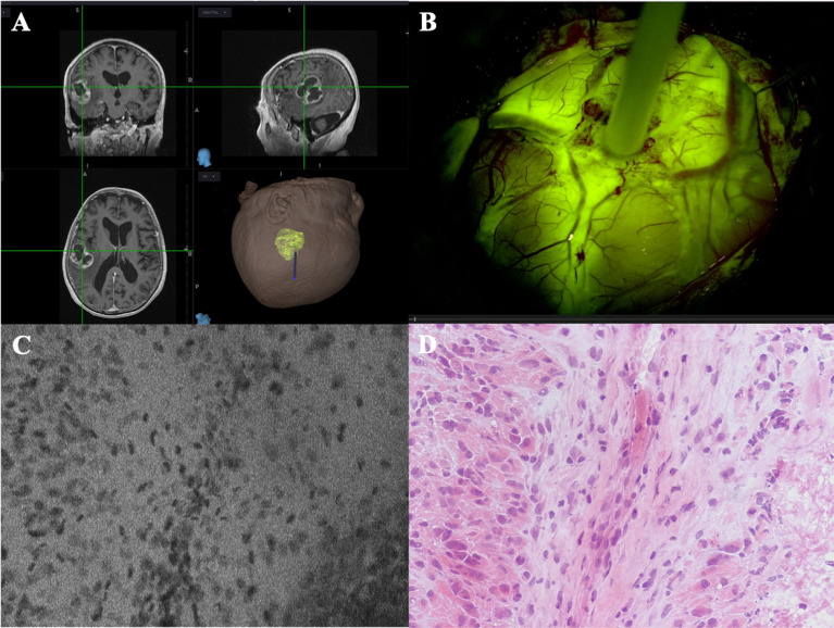
Figure 3.
Case example of an in vivo GBM case analyzed with CONVIVO (courtesy of Dr. Acerbi and Dr. Pollo, Fondazione IRCCS Istituto Neurologico Carlo Besta, Milan, Italy). (A) MRI preoperative images of a left parieto-temporal GBM, loaded on Stealth S8 navigation system (Medtronic, Minneapolis, USA). (B) CONVIVO stylet placed upon the center of the tumor, on the cerebral surface. As it can be seen, the tumor intensely enhances after intravenous SF administration. (C, D) CONVIVO and histological images of the point where the optical biopsy with CONVIVO was obtained. Disordered groups of dark nuclei cells can be seen, along with a stromal component among them. A low fluorescence area on CONVIVO, as it occurs in necrotic parts of the tumor, can be seen in the bottom right of panel (C), with its histological counterpart in the bottom right of panel (D).

