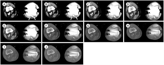
Fig. 13. Lower-extremity CT angiography images using the MEI plus technique of a 61-year-old male (body mass index, 18.5 kg/m2; CT volume dose index, 7.54 mGy; dose-length product, 939 mGy·cm) with peripheral arterial disease.
A-E. MEIs shown include (A) 40 keV, (B) 50 keV, (C) 60 keV, (D) 70 keV, and (E) 80 keV. Metal artifacts caused by the internal fixation material on the left femur and tibia affect the left popliteal artery evaluation. In the 40–50 keV images, the image quality is poor due to severe artifacts (arrows). In the 60–80 keV images, the image quality is adequate with slight metal artifacts. The metal artifacts gradually decrease in the images from 40 to 80 keV.
F-J. MEIs shown include (F) 40 keV, (G) 50 keV, (H) 60 keV, (I) 70 keV, and (J) 80 keV with optimized window settings (window level, 700 HU; window width, 3000 HU) due to the decrease in metal artifacts. With the window level and width adjustments, the vessel image quality is excellent without metal artifacts.
HU = Hounsfield units, MEI = monoenergetic image

