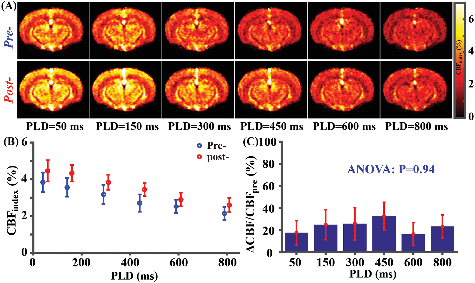
Figure 4.

CVR mapping results (N=4) with multi-PLD pCASL. (A) Average pre- and post-injection CBF weighted images at different PLDs. (B) Whole-brain CBF weighted signal for pre- and post-injection state at different PLDs. A small horizontal displacement between pre- (blue) and post-data (red) was added for display purposes. (C) Whole-brain CBF change (ΔCBF/CBFpre) at different PLDs. Error bars denote standard errors across mice.
