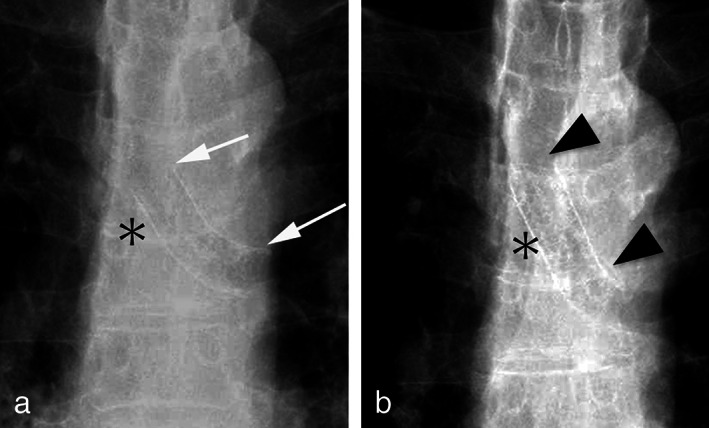
FIGURE 2.

(a) Chest radiograph showing the self‐expandable metallic stent placed in the left main bronchus (arrows). (b) Chest radiograph showing the dislocation of a self‐expandable stent to the oral side (arrowheads). The location of the carina is shown by an asterisk.
