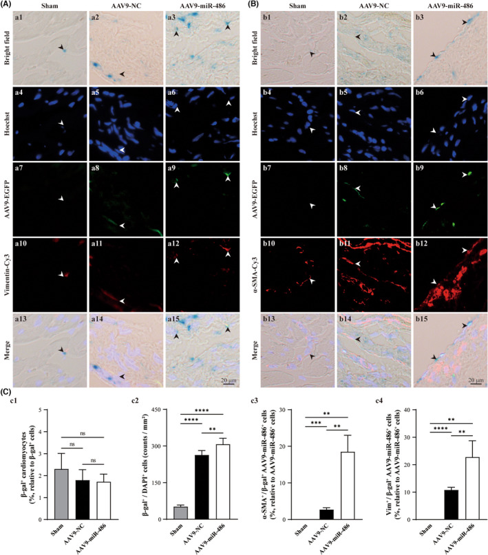
FIGURE 6.

miR‐486 overexpression increases the cellular senescence of CMFs in the infarct zone but not cardiomyocytes in the post‐MI myocardium in vivo. (A) Representative images of β‐gal staining (blue under bright field), anti‐Vimentin (Cy3; red under fluorescence) staining and Hoechst staining (blue under fluorescence), which were conducted in the same AAV9‐miR‐486‐EGFP (green)‐transfected myocardium sections at 8 weeks post‐MI. (B) Representative images of β‐gal staining (blue under bright field), anti‐α‐SMA (Cy3; red under fluorescence) staining and Hoechst staining (blue under fluorescence), which were conducted in the same AAV9‐miR‐486‐EGFP (green)‐transfected myocardium sections at 8 weeks post‐MI. β‐gal‐positive staining was performed to label senescent cells. Vimentin was used as a marker for CFs and CMFs. Vimentin‐ and α‐SMA‐positive cells were used to identify CMFs. Hoechst was applied to identify the nucleus. (C‐C1) Semiquantification of β‐gal+/cTnI+ cardiomyocytes in cross sections of whole myocardium for senescent cardiomyocytes. (C‐C2) Semiquantification of β‐gal+/DAPI+ noncardiomyocytes in the infarct zone. (C‐C3) Semiquantification of α‐SMA+/β‐gal+/AAV9‐miR‐486‐EGFP+ cells vs. AAV9‐miR‐486‐EGFP+ cells (%) in the infarct zone to analyse miR‐486 overexpression‐induced senescent CMFs. (C‐C4) Semiquantitative analysis of Vimentin+/β‐gal+/AAV9‐miR‐486‐EGFP+ cells vs. AAV9‐miR‐486‐EGFP+ cells (%) in the infarct zone to analyse miR‐486 overexpression‐induced senescent CFs and CMFs. n = 3.
