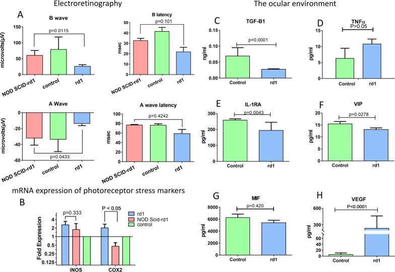
Fig. 2.
Retinitis pigmentosa animal models. Electroretinography was performed on immune competent rd1, NOD SCID-rd1 and control C57BL/6. A wave indicates the photoreceptor function while B wave indicates secondary retinal cell function responsible for signal transduction. In A, A wave functions displayed significant trends of vision conservation in NOD SCID-rd1 and the B wave function was conserved in NOD SCID-rd1 against rd1 and control at 4 weeks. The mRNA expression profile photoreceptor stress markers, iNOS and COX2, indicated that rd1 retina experienced higher levels of trauma in rd1 due to stress as compared to NOD SCID-rd1 and healthy retinas (B). The ocular environment of rd1 and control was studied which indicated that ocular environment can become pro-inflammatory due to TGF-B1 impairment as the TGF-B1 was significantly low in rd1 (C) while inflammatory cytokine TNFα level was increased in rd1 ocular milieu (D). Immune suppressive intra-ocular molecules IL-1RA (E) and VIP (F) and MIF levels (G) were decreased in rd1 ocular environment. This can potentially cause immune active T cells and monocytes to elicit potent responses. Elevated level of intra-ocular VEGF was detected in rd1 (H) which is an indication of ocular inflammation and pathogenic angiogenisis. n = 5

