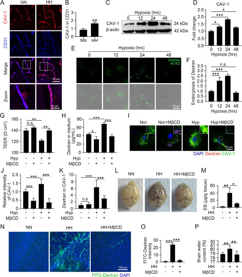
Fig. 3.
Hypoxia enhanced endothelial permeability through the upregulation of CAV-1-mediated endocytosis and triggered HACE. A and B C57BL/6 mice were exposed to HH (7600 m altitude) for 24 h. Brain slices were colabelled with CD31 and CAV-1 (A), and the fluorescence intensity of CAV-1 in the endothelium was counted (B) (**P < 0.01, n = 10). C to F bEnd.3 cells were exposed to 1% O2. The CAV-1 protein levels were detected by Western blot (C) and quantified (D). FITC-dextran was incubated with hypoxia-treated cells for 30 min (E), and the level of endocytosed dextran was calculated by counting the intensity in cells (F) (* P < 0.05 and *** P < 0.001). G to K bEnd.3 cells were treated with hypoxia for 24 h and then incubated with 5 mM MβCD for 1 h. After 30 min of FITC-dextran treatment, cell permeability was detected by TEER (G) and leakage of 40 kD FITC-dextran (H). CAV-1 was labelled by immunofluorescence (I), the intensity of CAV-1 (J) and dextran colocalized with CAV-1 (K) were counted (*P < 0.05, **P < 0.01 and ***P < 0.001). L to P C57BL/6 mice injected with 300 mg/kg MβCD were exposed to HH for 24 h and then reinjected with MβCD, which circulated for 1 h. After injecting EB via the tail vein for 1 h, the infiltration of EB into the blood vessels and tissues was observed (L) and the residual EB content in the tissues was quantified by colorimetry (M). Mice were injected with 40 kD FITC-dextran through the tail vein for 15 min, and the dextran distribution in the cortex was observed (N). The dextran fluorescence intensity was counted (O). The brain water content was measured by the dry and wet weight method (P) (*P < 0.05, **P < 0.01 and ***P < 0.001, n = 7)

