In the article “Introduction of Ultra-High-Field MR Imaging in Infants: Preparations and Feasibility” (K.V. Annink, N.E. van der Aa, J. Dudink, et al. AJNR Am J Neuroradiol 2020;41:1532–37 10.3174/ajnr.A6702) Table 1 and Fig 3 contained errors. The correct table and figure are the following:
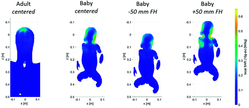
FIG 3.
Local SAR levels in adult head (left) and Charlie in the different coil positions. Shifts of an infant in the x and y directions are unlikely because of limited space; therefore, the results are not included in the figure. The SAR values when infant Charlie is positioned 50 mm in the x or y direction are comparable with those in the +50-mm FH position.
| Duke Centered | Ella Centered | Charlie Centered | Charlie –50 mm FH | Charlie +50 mm FH | |
|---|---|---|---|---|---|
| Global SAR | |||||
| Average SAR for 1 W input power (W/kg) | 0.066 | 0.069 | 0.091 | 0.075 | 0.109 |
| Average SAR per B12 (W/kg/μT2) | 0.462 | 0.465 | 0.320 | 0.477 | 0.484 |
| Average B1+ in central section for 1 W input power (μT)a | 0.379 | 0.385 | 0.542 | 0.397 | 0.475 |
| Peak SAR | |||||
| Peak local SAR (10 g averaged) for 1 W input power (W/kg) | 0.435 | 0.398 | 0.487 | 0.345 | 0.643 |
| Peak local SAR (10 g averaged) per B12 (W/kg/μT2) | 3.04 | 2.63 | 1.72 | 2.19 | 2.85 |
The power optimization procedure of the MR imaging scanner software calibrates the needed input power to achieve a certain B1 in the subject. This calibration is based on the average B1+ in a central section of the subject (brain in this case).
Textual Changes
ABSTRACT
BACKGROUND AND PURPOSE:
Cerebral MR imaging in infants is usually performed with a field strength of up to 3T. In adults, a growing number of studies have shown added diagnostic value of 7T MR imaging. 7T MR imaging might be of additional value in infants with unexplained seizures, for example. The aim of this study was to investigate the feasibility of 7T MR imaging in infants. We provide information about the safety preparations and show the first MR images of infants at 7T.
MATERIALS AND METHODS:
Specific absorption rate levels during 7T were simulated in Sim4life by using infant and adult models. A newly developed acoustic hood was used to guarantee hearing protection. Acoustic noise damping of this hood was measured and compared with the 3T Nordell hood and no hood. In this prospective pilot study, clinically stable infants, between term-equivalent age and the corrected age of 3 months, underwent 7T MR imaging immediately after their standard 3T MR imaging. The 7T scan protocols were developed and optimized while scanning this cohort.
RESULTS:
Global and peak specific absorption rate levels in the infant model in the centered position and 50-mm feet direction did not exceed the values used by the scanner to calculate energy deposition. Hearing protection was guaranteed with the new hood. Twelve infants were scanned. No MR imaging-related adverse events occurred. It was feasible to obtain good-quality imaging at 7T for MRA, MRV, SWI, single-shot T2WI, and MR spectroscopy. T1WI had lower quality at 7T.
CONCLUSIONS:
7T MR imaging is feasible in infants, and good-quality scans could be obtained.
RESULTS
Preparation: SAR Simulation
The global SAR and peak local SAR of the virtual infant model did exceed the SAR of the adult models (in the center position by +38% and +12% compared with Duke, respectively) (Table 1). However, the calculated SAR per B12 values are all below the values used by the scanner to calculate energy deposition (1.83 W/kg/uT2 for global SAR and 4.96 W/kg/uT2 for peak local SAR. Numbers as provided by the manufacturer).
Global and peak local SAR levels were highest when the infant model was positioned +50-mm FH. Furthermore, in this position, the local SAR showed hotspots in the neck/shoulder transitions (Fig 3).
The SAR per B12 was lower in the infant model in the center position than in the adult models, meaning that less power is needed to reach the same B1.
DISCUSSION
We demonstrated that scanning infants in a 7T scanner is feasible and results in good-quality images. While optimization of the sequences is ongoing, we already demonstrated that some sequences showed more details compared with 3T MR imaging.
When the infant’s head was further in the coil than isocenter, SAR levels were highest. Thus, the center position of the infant in the coil is essential. Therefore, the position of the infant’s head was constrained in the coil, making it mechanically impossible to put the infant’s head farther in the coil than center position.


