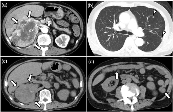
FIGURE 1.

Computed tomography (CT) findings. (a, b) First abdominal CT shows a 15 cm tumor in the right kidney with heterogeneous contrast in the arterial phase (arrows) with multiple lung metastases (arrowheads). (c) After three courses of immune checkpoint inhibitor therapy, CT showed that the primary tumor had decreased in size (arrows). (d) Three days after the end of the third course, CT shows wall thickening throughout the colon (arrows)
