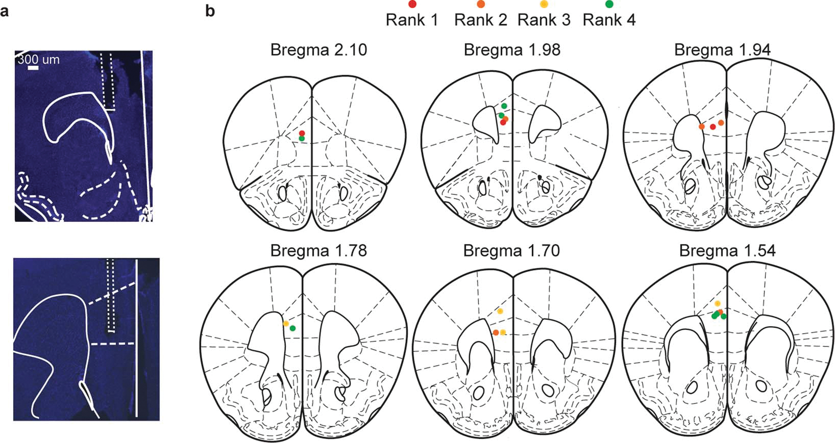
Extended Data Figure 4: Histological validation of electrode placements.

a, Representative images showing electrode track and lesions of mPFC electrode wires. b, Location of center for electrode lesions for all mice color coded by absolute rank across animals.
