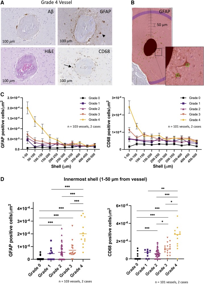
Figure 3.
Perivascular inflammation is observed predominantly around advanced grade vessels. (A) Aβ, H&E, GFAP and CD68 stains of a representative Grade 4 vessel. Arrowhead and arrow point to examples of a reactive astrocyte and activated microglial cell respectively. (B) Example of Sholl analysis within the MeVisLab software environment for GFAP-positive cells surrounding a vessel. Masked vessel from A is shown with successive 50 µm shells and GFAP-positive cells manually annotated (square markers). Inset displays two marked GFAP-positive cells. (C) Density of GFAP-positive (left) and CD68-positive (right) cells in successive shells surrounding vessels from two targeted cases with CAA (mean ± SEM). (D) Density of GFAP-positive (left) and CD68-positive (right) cells in innermost shell (1–50 µm from vessel) shown for each vessel analysed (median shown). *P < 0.05, **P < 0.01, ***P < 0.005 (after Bonferroni correction). Kruskal–Wallis test, P < 0.0001 for both GFAP- and CD68-positive cells. Post hoc pairwise comparisons shown using Mann–Whitney U-test with Bonferroni P-value adjustment. Included in GFAP analysis, number of vessels (Cases 1 and 2): Grade 0 = 11, 8; Grade 1 = 2, 7; Grade 2 = 24, 22; Grade 3 = 8, 9; Grade 4 = 10, 2. Included in CD68 analysis, number of vessels (Cases 1 and 2): Grade 0 = 10, 8; Grade 1 = 1, 7; Grade 2 = 24, 22; Grade 3 = 8, 9; Grade 4 = 10, 2.

